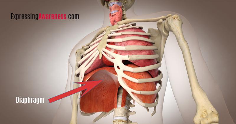
What is the diaphragm?
Diaphram is a large dome-shaped muscle that continuously contracts and relaxes in a rhythmic pattern matching your breath. The same as your breathing, diaphragm actions are often involuntary, and you can exert voluntary control over it for a brief time.
Where is the diaphragm located?
Diaphram is located below your lungs, separating your lungs and heart from your stomach.
What is the function of the diaphragm?
Diaphram muscles serve two primary purposes.
Respiratory
Diaphram muscle’s primary role is respiration, which is the process of breathing.
Diaphragm contracts and flattens as you inhale, creating suction for the lungs to expand and pull the air in. Understanding this suction activity helps you improve your breathing during exercise.
The feeling that you are forcing the air in during a deep exhalation is just an illusion. In reality, you are creating a suction that pulls the air in.
The stronger the suction, the more air gets in faster. The stronger your diaphragm muscle is, the more suction it can create. The weaker the diaphragm muscle is, the more challenging time you have with taking deep breaths.
Nonrepiratory
Diaphragm muscle serves nonrespiratory functions as well. Increasing the abdominal pressure helps the body get rid of vomit, urine, and feces.
Increased abdominal pressure also stabilizes the spine during heavy lifting. Valsalva maneuver is an example of using abdominal pressure to stabilize the core. Valsalva maneuver refers to holding your breath for a very brief period during heavy lifting.
The lower esophageal sphincter (LES) is a band of muscle that helps prevent reflux which happens when stomach contents come back up into the throat.
Diaphragm muscle assists LES to prevent acid reflux. A hiatal hernia may decrease the sphincter pressure necessary to maintain the anti-reflux barrier.
What nerve innervates the diaphragm?
The phrenic nerve controls the diaphragm. The phrenic nerve goes from the neck to the diaphragm and is responsible for breathing movements.
Diaphragm Anatomy
Aortic opening, caval opening, and esophageal opening allow vital anatomical structures to pass through the diaphragm, connecting the stomach organs to those within the chest cavity.
- Aortic opening. The main artery (aorta) transporting blood from the heart passes through this opening. The main vessel for the lymphatic system (thoracic duct) also passes through the aortic opening.
- Esophageal opening. Both the vagus nerve responsible for parasympathetic nervous system activation and the esophagus pass through the Esophageal opening.
- Caval opening.The large vein ( inferior vena cava) transports the blood from the heart passes through the caval opening.
Watch the video below for an effective diaphragm breathing exercise.


